


Arthrosis is a chronic pathology that affects the connective tissue structures of the musculoskeletal system.The disease is characterized by a progressive course with gradual destruction of cartilage tissue.Arthrosis is detected in most patients after 65 years of age, because one of the reasons for its development is the natural aging of the body.
The appearance of degenerative-dystrophic pathology is caused by previous injuries, endocrine and inflammatory diseases, excessive physical activity or, on the contrary, a sedentary lifestyle.The leading symptoms of arthrosis are joint pain, swelling and limitation of movement.
To diagnose the pathology, instrumental studies are carried out - radiography, arthroscopy, MRI, CT.Arthrosis of the 1st and 2nd severity is treated conservatively with drugs, physiotherapeutic and massage procedures and exercise.In case of irreversible destructive changes in the joints, surgical intervention is indicated - arthrodesis, endoprosthetics.
Pathogenetic mechanisms
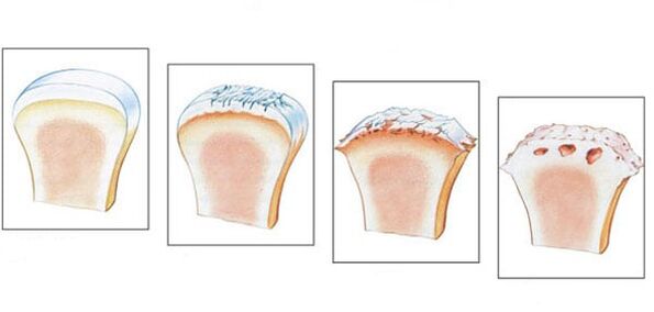
In arthrosis, pronounced changes occur in the internal structures of the connective tissue.Deforming erosions occur on cartilage tissues, which causes the destruction of collagen fibers, as well as proteoglycans, which consist of proteins (5-10%) and glycosaminoglycans (90-95%).As a result, the collagen network loses stability and metalloproteinases begin to be released, destroying all types of extracellular matrix proteins.Destruction is accelerated by increasing the biosynthesis of collagenases and stromelysin.Typically, normal quantitative enzyme values are controlled by cytokines - small peptide information molecules.But as arthrosis progresses, the concentration of these proteins decreases, which causes the release of a large number of enzymes that damage cartilage tissue.
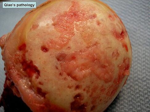
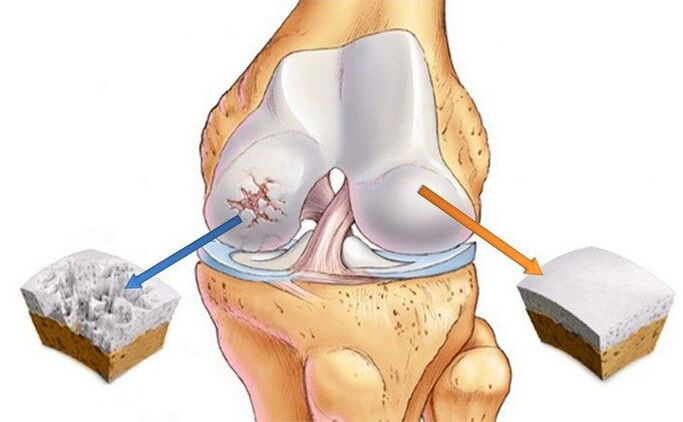
Proteoglycans with a changed structure begin to absorb water molecules that they are unable to retain.Because of this, excess fluid enters the collagen fibers.They "swell" and lose strength and elasticity.Negative changes occur in both the qualitative and quantitative composition of the synovial fluid.In arthrosis, the concentration of hyaluronan in it decreases.Hyaline cartilages no longer receive enough nutrients and oxygen for their regeneration.Softening foci are formed in the cartilage tissue, and then cracks and specific necrotic growths appear.The heads of the bones become exposed and begin to undergo microtrauma when they move relative to each other.
Causes and provoking factors
The reasons for the development of primary (idiopathic) arthrosis have not yet been determined.It occurs in the absence of any provoking factors, therefore theories are put forward about a hereditary predisposition to premature destruction of cartilage.Secondary arthrosis occurs as a result of other joint pathologies or previous injuries.What can cause degenerative-dystrophic disease:
- injury to the joint or nearby connective tissue structures - fracture, dislocation, damage to the meniscus, partial rupture of muscles, ligaments, tendons or their complete separation from the bone base;
- congenital dysplastic joint development disorder;
- disruption of endocrine glands, disruption of metabolic processes;
- rheumatism or rheumatic fever;
- rheumatoid, reactive, metabolic, psoriatic or gouty arthritis, polyarthritis;
- purulent arthritis caused by streptococcus, epidermal or Staphylococcus aureus;
- tuberculosis of any localization, brucellosis, chlamydia, gonorrhea, syphilis;
- degenerative diseases, for example, osteochondritis dissecans.
Hypermobility of the joints, caused by the production of special collagen, predisposes to the occurrence of arthrosis.This condition is detected in 10% of the planet's inhabitants and is not considered a pathology.But hypermobility is accompanied by weakness of the tendon-ligament apparatus, which leads to frequent injuries, especially of the ankle joint (sprains and ruptures of ligaments, dislocations).
Osteoarthritis is sometimes caused by hematopoietic disorders, such as hemophilia.Hemarthrosis, i.e. bleeding into the joint cavity, causes deterioration of cartilage trophism and its destruction.
Predisposing factors are age, frequent joint loads that exceed the limits of their strength, excess weight, surgical interventions and hypothermia.

The risk group includes women during menopause, people living in unfavorable environmental conditions or in contact with toxic chemical compounds.If there is a lack of foods with vitamins and microelements in the diet, prerequisites are created for the gradual destruction of hyaline cartilage.
Clinical picture
The danger of arthrosis lies in the absence of symptoms in the first phase of its development.Pathology is clinically manifested gradually, the first signs appear against the background of significant destruction of cartilage tissue.At first, a person feels a slight pain that does not have a clear localization.Appears after physical activity - weight lifting, sports training.Sometimes the first clinical manifestation is a creaking or clicking sound when flexing or extending the joint.The person begins to notice that some movements are difficult.However, in the initial stage of arthrosis, stiffness occurs in the morning and soon disappears.
As the disease progresses, the pain is also felt at night, causing not only sleep disturbance, but also the appearance of chronic fatigue.The severity of the pain syndrome in the second phase increases with changes in weather, worsening of chronic pathologies and acute respiratory viral infections.Range of motion is noticeably reduced.The cause of the stiffness is the thinning of the cartilage, as well as the conscious restriction of the person's movements in an attempt to avoid pain.This leads to increased stress on the opposite joint, which causes further damage.Arthrosis is also characterized by other specific symptoms:
- pain causes spasms of skeletal muscles and the development of muscle contractures (restriction of passive movements in the joint);
- crunching in the joints, clicking, crackling when moving become constant, occur with almost every movement of the bones in relation to each other;
- painful muscle spasms often occur;
- the joints are deformed, which leads to posture and gait disorders;
- in the third stage of arthrosis, the deformation is so pronounced that the joints are bent, and the range of motion in them is significantly reduced or completely absent;
- with third-degree arthrosis of the knee, ankle joint, hip joint, the patient uses a stick or crutches when moving.
In the absence of treatment, the pathology progresses, and during its course, remissions are replaced by relapses, and the frequency of exacerbations constantly increases.Stiffness in movements in the morning now does not disappear for a long time, it becomes permanent.
When examining a patient with stage 1 arthrosis, the doctor notices only slight swelling of the joint and complete preservation of the range of motion.In pathology of the 2nd degree, palpation reveals pain and slight deformity.Bone thickening is observed in the area of the joint space.
Arthrosis is characterized by the development of synovitis - inflammatory processes in the synovial membranes of the hip, knee, ankle and shoulder joints.Their leading symptom is the formation of a rounded seal in the area of the joint, when pressure is applied to which fluid movement (fluctuation) is felt.Acute synovitis can be accompanied by an increase in temperature up to 37-38 °C, headache and indigestion.
Diagnostics
The diagnosis is made on the basis of the results of instrumental studies, characteristics of the clinical picture, anamnesis and complaints of patients.A general blood and urine test is not very informative - all values remain within normal limits if arthrosis is not caused by metabolic pathology.With the development of synovitis, the rate of erythrocyte sedimentation increases (30 mm/hour), and the level of leukocytes and fibrinogen in the blood also increases.This indicates an acute or chronic inflammatory process taking place in the body.Changes in biochemical and immunological parameters occur in secondary forms of arthrosis.
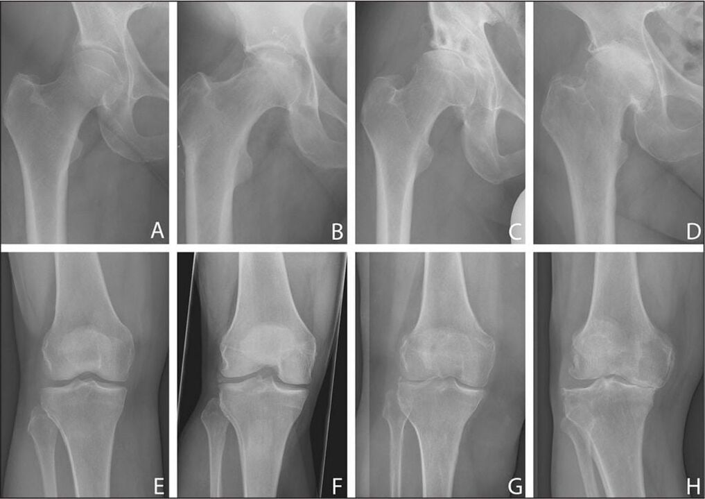
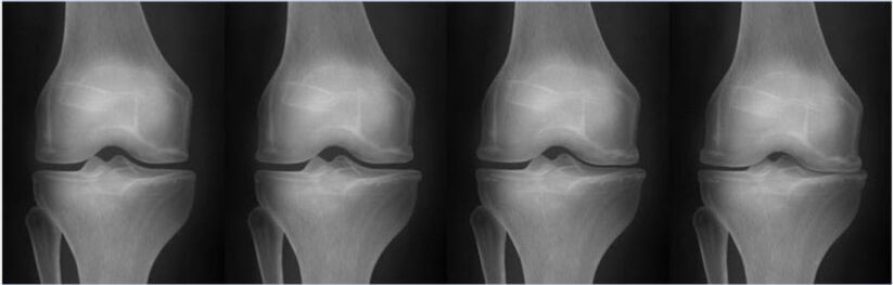
The most informative method for diagnosing degenerative-dystrophic pathology is radiography in frontal and lateral projection.
| Stages of arthrosis according to the Kellgren-Lawrence classification (1957) | X-ray signs of pathology |
|---|---|
| Initial | No radiological signs |
| First | Unclear, uneven narrowing of the joint space.Slight flattening of the edges of bone plates, formation of initial osteophytes or their absence |
| Second | Pronounced narrowing of the joint space, 2-3 times larger than normal, formation of a large number of osteophytes, subchondral osteosclerosis.The appearance of cystic clearings in the epiphyses |
| Third | Appearance of pronounced subchondral osteosclerosis and large marginal osteophytes, significant narrowing of the joint space |
| Fourth | Formation of coarse massive osteophytes, almost complete fusion of the joint space, deformation and compaction of the epiphyses of the bones that form the joint |
If, after studying the X-rays, the doctor doubts the diagnosis, a CT scan is prescribed.And in order to assess the condition of the connective tissue structures located near the joint, an MRI is performed.By using contrast, it is possible to dynamically assess blood flow to tissues and determine the stage of the inflammatory process during the development of synovitis.
Basic methods of therapy
Arthrosis is still an incurable disease, because there are no pharmacological drugs for cartilage tissue regeneration.The main goal of therapy is to prevent the progression of the pathology and maintain joint mobility.The treatment is long-term, complex, using local and systemic drugs.Patients should avoid strong stress on the joint and, if necessary, limit the range of motion with orthopedic aids - orthoses, elastic bandages.Overweight patients must adjust their diet to gradually reduce body weight and follow a diet.
After achieving stable remission, patients are shown daily physical therapy exercises.The first trainings are performed under the guidance of a physical therapy doctor, then the patient performs a set of exercises at home.Exercise therapy can be supplemented with swimming, yoga and cycling.
To reduce the intensity of pain, drugs from different clinical and pharmacological groups are prescribed:
- non-steroidal anti-inflammatory drugs in the form of ointments, tablets, solutions for parenteral administration with active ingredients;
- joint injections of anesthetic solutions in combination with glucocorticosteroids;
- muscle relaxants to eliminate muscle spasms and restrictive contractures.
Therapeutic regimens include B vitamins, sedatives and, if necessary, tranquilizers and antidepressants.Long-term use requires chondroprotectors.This is the only group of drugs that have the ability to partially restore cartilage tissue.
To increase their clinical activity, physiotherapeutic procedures are carried out - laser therapy, magnetic fields, UHF therapy.
Any pain in the joints should be a signal for an urgent consultation with a doctor.Therapy carried out in the initial stage of arthrosis will stop the destruction of cartilage and avoid loss of performance and disability.